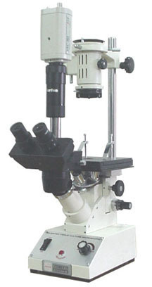
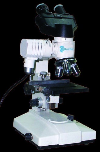
About Trinocular Inverted Microscope
This Latest model has been designed to examine and sturdy tissue cultures, bacteria plankton, Protozoan and similar Micro-organisms in media. Sturdy base with built in 6V-30W continuously variable light intensity control. Stage size180x150mm with extension plates for large bottles & micro filter work with detachable or co-axial mechanical graduated stage. Illumination Koehler system provided by 6V-20W. Binocular Observation head has been provided with coated prismatic system. Latest co-axial coarse & fine focusing system based on a4- gear reduction system traveling on ball bearing guides with highly sensitivity fine motion with a graduation reading to 0.002mm.
Quadruplenose piece revolving turret to carry objectives:
Objectives : 5x, 10x, 20x & 40x
Eyepieces : WF 10x (Pair) with eye guards.
Filters : Blue & White.
Specification as model BTM 417 but provided witha Trinocular head with binocular observation and straight tube forPhotomicrography
Sturdy Design for Reliable PerformanceThe microscope rests on a robust anti-vibration cast metal base, ensuring stability during detailed sample observation. Its mechanical stage offers X-Y movement with fine adjustments, while the ergonomic inclined eyepiece tube provides comfortable, ambidextrous viewing for prolonged usage.
Advanced Imaging and Camera CompatibilityDesigned to integrate seamlessly with most C-mount digital and CCD cameras, this microscope enables high-resolution imaging and video capture. Users can connect cameras easily via the phototube, capturing images in JPEG, BMP, or TIFF formats and recording videos up to 1080p.
Precise Optics for Cell Culture StudiesWith long working distance objectives and anti-fungus treated achromatic lenses, the microscope delivers sharp images ideal for observing living cells and tissues. Fine and coarse coaxial focusing systems allow accurate adjustments, making it perfect for research, academic, and clinical applications.
FAQ's of Trinocular Inverted Microscope:
Q: How does the trinocular design benefit laboratory applications?
A: The trinocular head allows simultaneous viewing through both eyepieces and the phototube, enabling direct attachment of a camera for capturing images or live video during demonstrations or analyses, which is highly beneficial for documentation and sharing results in research environments.
Q: What is the process for attaching a digital camera to the microscope?
A: To attach a digital or CCD camera, simply use the C-mount adapter on the phototube of the trinocular head. Ensure the camera is compatible for integration, and follow the manufacturer's guidelines for secure connection and optimal imaging performance.
Q: Where is this microscope typically used, and for what research purposes?
A: This inverted microscope is commonly utilized in laboratories, hospitals, and research facilities for observing living cells, tissues, and samples in culture media. It is widely employed in cell biology, microbiology, and tissue engineering studies.
Q: What are the benefits of the anti-fungus treated objectives?
A: Anti-fungus treated achromatic objectives keep optical performance high and prevent damage from humidity, ensuring clear, reliable images and extending the lifespan of the microscope, especially in environments prone to fungal growth.
Q: How can the stage movement range improve the usage experience?
A: The mechanical stage offers an X-Y movement range of 75 mm x 50 mm, along with a vernier scale for precise sample positioning. This facilitates accurate observation of specific areas within samples, enhancing efficiency during cell culture studies.
Q: When should the illumination source be switched between LED or halogen?
A: Switch between LED and halogen illumination depending on sample thickness and desired image clarity. LEDs offer adjustable brightness and longer life, while halogen bulbs provide intense, warmer light ideal for brightfield applications or thicker specimens.
Q: What advantages does the fine focus division of 0.002 mm provide?
A: The ultra-precise fine focus division allows detailed focusing adjustments, enabling users to resolve minute cellular structures and optimize imaging quality, which is crucial for tasks requiring high accuracy, such as cell culture analysis and microstructure examination.


 English
 Spanish
 French
 German
 Italian
 Chinese (Simplified)
 Japanese
 Korean
 Arabic
 Portuguese
 Send Inquiry
 Get Latest Price
